Corporate news
24-11-2025
Publication date: 26-09-2024
Updated on: 17-10-2024
Topic: Corporate news
Estimated reading time: 1 min
Medical Editor
Antonio EspositoEditor and Translator
Anastasiia ByvaltcevaThe vehicle fleet of Ospedale San Raffaele is expanding, welcoming two of the most advanced and innovative technologies on the market: the Photon counting CT and a state-of-the-art 3 Tesla MRI.
Located within the Unit of Advanced Imaging for Personalized Medicine - AI4PM, directed by Professor Antonio Esposito, deputy scientific director and full professor of Imaging Diagnostics and Radiotherapy at the Faculty of Medicine and Surgery at Università Vita-Salute San Raffaele, the new machines will be used:
Thanks to the use of the most sophisticated imaging equipment, characterized by unique technologies and specific Artificial Intelligence algorithms, the imaging diagnostics at Ospedale San Raffaele opens its doors to the latest frontiers of advanced diagnostic technology, reaffirming itself as a cutting-edge Research and Care Institute at the forefront of new diagnostic-therapeutic horizons for its patients.
In this regard, Professor Esposito offers an overview of the new machines, explaining their features and benefits.
“This scanner represents the first FDA-approved and CE-marked CT technology in the world equipped with a photon counting detector,” explains Prof. Esposito.
“All other CT scanners are equipped with conventional ‘energy integrating detectors,’ which work by integrating the detection of thousands of photons and converting them into a single light signal, which is then transformed into an electrical signal. The photon counting detector of our scanner directly converts the X-ray photons into an electrical signal, which is then used to create the image.”
In photon counting technology, the energy of each individual photon is measured and converted into a single electrical signal proportional to the photon's energy. This results in significant advantages:
Additionally, within the scanner, the ultra-high spatial resolution and spectral resolution of the images are combined with the highest temporal resolution of any CT scanner on the market, allowing the visualization of extremely precise details even for structures that naturally move due to the heartbeat or breathing, providing greater diagnostic reliability.
These features make it a unique CT instrument with unparalleled performance compared to any other CT scanner, setting new standards in the diagnosis of diseases, with particular attention to oncological, cardiovascular, and pulmonary diseases. For example, it is literally possible to see tumors that were previously invisible or to view in detail the flow of blood inside coronary stents, which were inaccessible with conventional CT technology.
The technical possibilities offered by this new technology define it as a valuable clinical and research tool, and they inherently create the need to develop dedicated research programs aimed at exploring, determining, and validating new diagnostic and treatment protocols and processes.
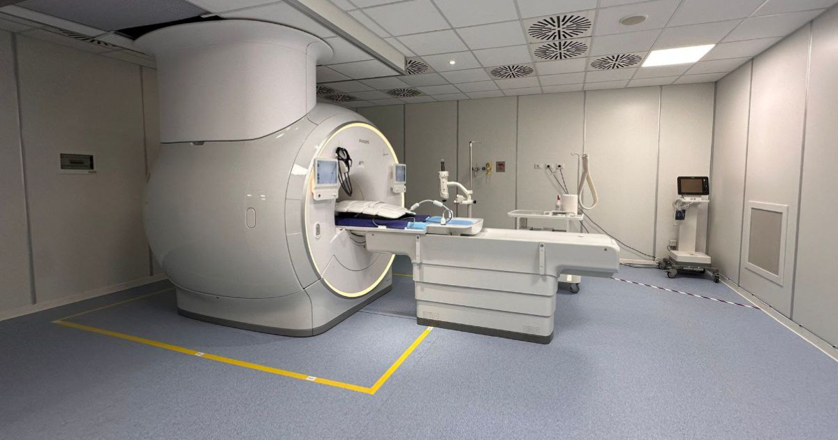
The new 3 Tesla Magnetic Resonance Imaging (MRI) machine at San Raffaele is an innovative non-invasive imaging diagnostic tool that allows the acquisition of ultra-high-resolution images, without using ionizing radiation or radioactive tracers, and often without the need to inject contrast agents.
"This new machine, equipped with high-performance XP gradients and artificial intelligence, offers excellent clinical performance and enables the management of complex research programs," explains Esposito. By combining high-performance hardware structures with the most advanced software development in MRI, it provides unprecedented image quality and advanced workflows, elevating diagnostics to new standards and completing exams in less time, benefiting:
The exceptional performance of this MRI scanner is particularly suited to supporting advanced precision diagnostics and clinical research protocols based on quantitative MRI biomarkers. This technology also serves as a valuable platform for designing, developing, and validating new imaging protocols, diagnostic and prognostic biomarkers, as well as new personalized diagnostic and treatment processes through dedicated research and development projects.
Additionally, the technical features of this scanner allow for exceptional quality in diffusion-weighted imaging, which accurately studies tissue cellularity, a factor correlated with tumor transformation. It is, therefore, possible to perform whole-body MRI studies for screening, diagnosis, and follow-up, characterized by extreme diagnostic accuracy and precision.
"The whole-body MRI exam lasts about 35/40 minutes," continues Esposito, "scanning the entire body from the head to just above the knees, and acquiring multiparametric images with very high sensitivity for oncological alterations, even in very early stages, as well as for vascular alterations of major vessels, even before they become symptomatic. Specific examples include:
The same quality and benefits are also guaranteed in MRI exams of specific anatomical regions, such as the heart, all abdominal and pelvic organs (both male and female), and joints."