What are contrast agents and what are they used for?

出版日期: 24-07-2023
更新日期: 24-07-2023
主题: Radiology
预计阅读时间: 1 分钟
文章作者
Lara Benvenuti
编辑和译员
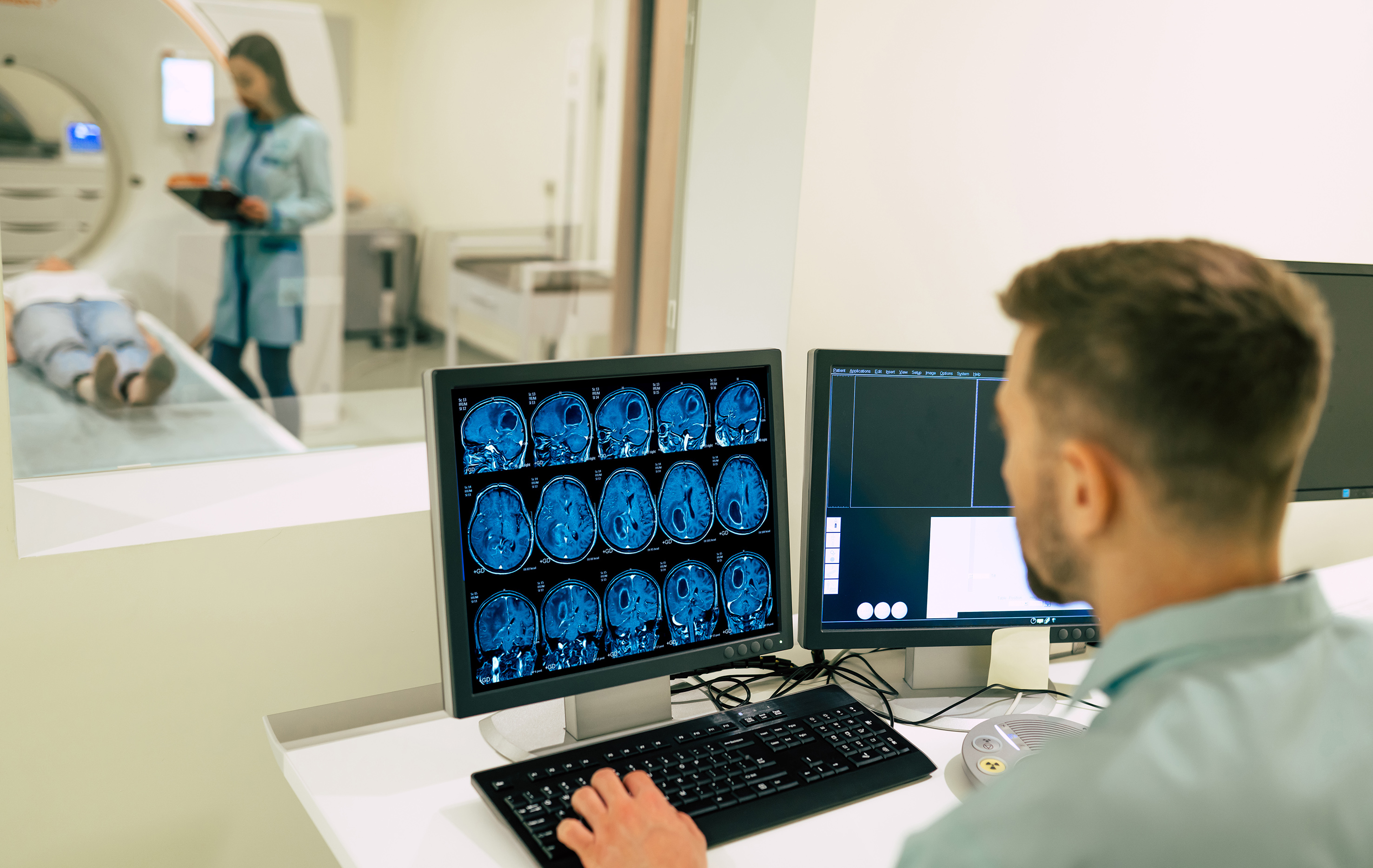
Viktoryia LuhakovaContrast media (mdc), or contrast fluids, are substances used in diagnostic imaging that help already high-resolution examinations such as, for example, CT and MRI, to improve image quality by bringing out tissue details and possible lesions that would not otherwise be visible.
Contrast media, which can be of different types depending on the type of examination with which they are associated, have different characteristics, and before administering them, it is necessary to understand what the possible risks to the patient might be and how to deal with them.
We discuss this with Dr. Gaetano Ruffo, head of the Radiology and Diagnostic Imaging Service at Istituto Clinico San Siro.
Contrast media in CT scanning
“One of the imaging tools that makes use of radiological contrast fluids is undoubtedly CT,” Dr. Ruffo explains. “In this case, an organo-iodine contrast agent is used.
In the present day, the evolution of contrast agents has led to drugs that are more tolerable to the body and normally well accepted even by highly allergic patients. It must be taken into account that contrast agents are macro-molecules, that they are not routinely used drugs of which intolerance is known and that they may result in adverse reactions that cannot be predicted at the time of administration.
For already known allergic patients, preparation for the examination is carried out (prophylaxis by taking cortisone and antihistamines), while, in specific cases, when reactions to contrast media such as anaphylactic shock are already known, the subject is refused this type of examination. In any case, when mdc administration is performed, the availability of the anesthesiologist is expected for any adverse reactions. An informed consent form is given and explained to the patient and a consent signature is requested”.
How to prepare for a CT scan with contrast medium
The organo-iodine contrast agent used in CT can cause damage to the body with toxicity mainly on the renal tubule, so good renal function is required on the part of the patient. In particular, it is necessary to undergo a preliminary blood test (plasma creatinine) for taking contrast medium. The radiologist will assess whether there are contraindications, and it is up to him or her to decide whether or not to perform the examination with contrast medium.
“Nowadays”, he continues, “reactions to contrast medium are rare. Obviously, they can range from mild to severe reactions that are potentially dangerous to the patient's health. Contrast medium injection is performed by the radiologist, with the availability of the Anesthesiology and Resuscitation Service for the occurrence of any complications”.
Contrast media in MRI
Contrast media are also used in MRI: these are no longer organo-iodine media as in CT, but mainly contrast media containing gadolinium, an element in the periodic table of elements that is part of the non-metals – rare earths, which is well tolerated by the body and has almost no side effects.
Contrast medium for MRI is also excreted renally, and therefore the same precautions apply as for organo-iodine contrast medium, i.e., kidney function is assessed with plasma creatinine prior to the examination.
Among the gadolinium contrast agents, some have precisely a specific target organ: in particular, there are the hepato-specific ones i.e., those that allow the detection of liver lesions, which tend to contrast mainly hepatic (i.e., liver) tissue.
Other uses of gadolinium contrast medium
“One of the major uses of contrast medium nowadays is in the Multiparametric MRI of the prostate, an examination called the golden standard for figuring out whether a nodule is benign or malignant, and one of the parameters is precisely the contrast medium,” he continues. “It is becoming, for this very reason, an irreplaceable examination in urology.
Other indications are, for example, the evaluation of abdominal masses suspected to be neoplastic or in the case of surgery on a muscle to find out whether or not it is a sarcoma (malignant tumor) or a lipoma (benign tumor). They are also used to study the brain and central nervous system, including in degenerative diseases such as, for example, Multiple Sclerosis.
Some MRI examination sequences for the study of arterial or venous vascular districts (angio-RM) are performed with the injection of contrast medium.”
Joint contrast medium
There are MRI (and also CT) investigations that involve the injection of contrast medium directly into the joint cavities to highlight anatomical details that are difficult to study with examinations performed only under baseline conditions.
The most important example by frequency is that of arthroresonance (arthro-RM) of the shoulder for the study of joint instability. After episodes of dislocation between the head of the humerus and the glenoid labrum of the scapula, arthro-RM allows for study of the glenoid labrum of the scapula with high anatomic detail. This MRI is considered an important premise for eventual reconstructive surgical therapy.
Other uses of mdc include follow-up MRI investigations in patients operated on for intervertebral disc herniations; in this context, the contrast agent allows for differentiation of the possible presence of post-surgical scar material from recurrences of herniated pathology.
Disposal of contrast fluids
“With good renal function, both gadolinium and organo-iodine contrast agents are disposed of within hours to a maximum of one day, and this is considered by the literature to be a sufficient standard,” Ruffo concludes. “In the case of using hepatospecific contrast agents, disposal is by the hepatobiliary route, then by the digestive tract.
In conclusion, contrast agents are a very important and frequently used aid in diagnostic imaging. As with all medications, there are precautions and warnings to be observed in their use.”